Beware the slowly-growing mass - and metal artifact! A patient diagnosed with left pelvic osteosarcoma 4.5 years ago demonstrated good response to initial treatment and was being followed off-treatment with serial q3 month CT Chest and Pelvis exams.
Initial radiograph and T2 fat-saturated MR image demonstrating a permeative pelvic mass with large mineralized soft tissue component and aggressive periosteal reaction.
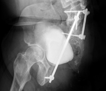
Subsequent radiograph demonstrating surgical changes of total hip disarticulation, resection of the left hemipelvis, and placement of metallic rod and strut graft between the lumbosacral spine and right pubis.
Earliest CT image in bottom left corner. A slow-growing, avidly-enhancing soft tissue mass was noted underlying the metal strut and largely obscured by in-plane streak artifact. Eventual growth necessitated tissue sampling; the pathology report confirmed high-grade recurrent osteosarcoma.
Osteosarcoma is the most common pediatric primary bone malignancy. Conventional high-grade is the most common of several sub-types, and occurs primarily at physeal sites with rapid growth (distal femur, proximal tibia, proximal humerus).
Factors affecting prognosis include: presence of metastatic disease at the time of diagnosis, response to chemotherapy, size and location of primary tumor (larger and central/axial are worse), age (outcomes in younger children are better), response to chemotherapy, and recurrence/relapse (longer disease-free intervals have better survival). Whether local recurrence affects overall survival is not known.