Infant with limp, diagnosed with an incomplete fracture of the tibia by an outside organization 3 months prior to presentation. After casting and follow-up imaging, the patient continued to have pain and limp, awaken at night, and lost 10 pounds. Inflammatory labs were normal.
![]() MR imaging shows smooth cortical thickening of the proximal tibial diaphysis, inflammation of the marrow and periosteum extending to soft tissues surrounding the bone.
MR imaging shows smooth cortical thickening of the proximal tibial diaphysis, inflammation of the marrow and periosteum extending to soft tissues surrounding the bone.
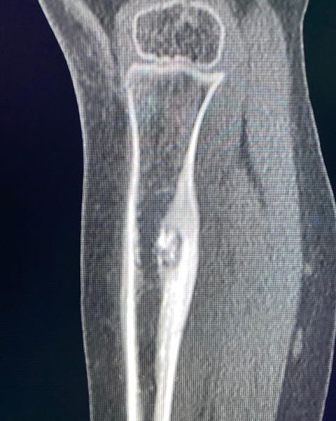
![]() CT imaging shows smooth cortical thickening posteriorly with a well-defined intra-cortical lucent lesion consistent with a nidus, confirming the diagnosis of Osteoid Osteoma.
CT imaging shows smooth cortical thickening posteriorly with a well-defined intra-cortical lucent lesion consistent with a nidus, confirming the diagnosis of Osteoid Osteoma.
![]() Osteoid osteoma is a benign non-neoplastic lesion most commonly occurring in the diaphysis or metaphysis of long bone and associated with dense fusiform reactive sclerosis.
Osteoid osteoma is a benign non-neoplastic lesion most commonly occurring in the diaphysis or metaphysis of long bone and associated with dense fusiform reactive sclerosis.
![]() Less common sites include subperiosteal, epiphyseal, and intra-articular locations; within flat bones (e.g. pelvis) and small bones (foot and hand); and the spine.
Less common sites include subperiosteal, epiphyseal, and intra-articular locations; within flat bones (e.g. pelvis) and small bones (foot and hand); and the spine.