Nightmare Endourology Case
By Thomas Johnson
A 51-year-old obese patient with spina bifida, epilepsy, left ventricular dysfunction and lifelong warfarin due to a deep vein thrombosis (DVT) had an ileal conduit stone noted as an incidental finding on a CT scan (Figure 1). A 6mm stone was present in the right lower pole. Her urine was colonised with pseudomonas and she was MRSA positive.
Under general anaesthesia, a nephroscope was used to fragment the stone in situ which appeared visually clear. This was a technically difficult procedure as the stone was freely mobile in the conduit.
Fragmentation was achieved but a postoperative CT demonstrated a significant residual burden (Figure 2).
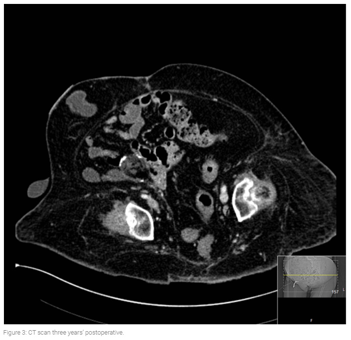
The patient was lost to short-term follow-up but a three-year CT scan demonstrated her urinary tract to be stone free (Figure 3).
Learning points
Ileal conduit stones are very rare.
Fragmentation is very time-consuming due to the mobility of the stone.
Large fragments and indeed upper tract fragments will drain via the conduit.