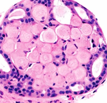
Collagenous Spherulosis:
This microphotographs is showing a dense spherules of eosinophilic basement membrane-like material surrounded by cells. This arrangement gives the proliferation a fenestrated or cribriform appearance.
Its an incidental microscopic finding in 1-2% of biopsies that contain hyperplastic ductal lesions. Important to recognize CS because lesion may superficially resemble cribriform DCIS.CS may also superficially resemble low-grade adenoid cystic carcinoma.
Spherules composed of varying amounts of basement membrane-like material, including polysaccharides, laminin, and type IV collagen
Positive staining for PAS and Alcian blue by histochemistry.In some cases, spherules contain mucoid-like material (“mucinous spherulosis”).Spherules surrounded by inner myoepithelial layer and outer luminal layer of cells.Myoepithelial cells may become attenuated and difficult to appreciate in H&E sections.Immunohistochemical stains for myoepithelial cells (calponin, p63, myosin heavy chain) to highlight myoepithelial cells may be helpful.Spherules and cellular arrangement give rise to appearance of cribriform or fenestrated proliferation when viewed at low power.