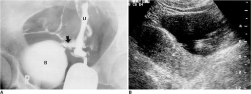
Figure 4: Vesicouterine fistula in a patient with urinary incontinence after a cesarean section.A. Hysterosalpingogram shows a fistulous tract (arrow) and contrast leakage from the uterine cavity into the bladder.B. Longitudinal ultrasonographic scan of the pelvis demonstrates hyperechoic line in continuity with the endometrium of the uterus andbladder (Reprinted, with permission, from reference 3) (B, bladder; U, uterus; arrows, fistula).
Mentions: Radiographically, both cystography and hysterography have been successfully used to demonstrate the presence of vesicouterine fistulas. In his review of published reports, Tancer found that hysterography was the most reliable diagnostic technique (7). Intravenous urography has been reported to demonstrate contrast medium entering the vagina but found to be of no value in distinguishing vesicovaginal and vesicouterine fistulas (8). Although fistulas are difficult to diagnose sonographically, the literature contains several reports on their ultrasonographic diagnosis. Park et al. reported that sonography can demonstrate the fistulous tract as double echogenic lines between the endometrium of the anterior wall of the uterine body and the mucosal layer of the posterior wall of the bladder (3) (Figs. 4, 5).