VIEWING MEDIASTINAL AND HILAR REGIONS :
Today I will be discussing about Mediastnum and Hilar abnormalities.
First of all before moving ahead my aproach is to locate and ensure that opacity is in mediastinum and then come which part of mediastinum and lastly the diagnosis.
A. So to diffentiate between pulmonary and mediastinum opacities -->
- Mediastinum opacities have smooth borders well defined and sharply denarcated
2.mediastinal opacities occupies generally the central portion of frontal x-rays.
3 no secondary signs can be visible like air bronchogram, cavitation or satellite nodule.
Pulmonary opacities has rugged margins secondary changes and generally unilateral lung
B. Which part of mediastinum it involves anterior posterior or middle
Few signs helps
-
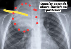
Cervicothoracic sign (upper mediastinum anterior or posterior)
-
Hilum convergence (middle mediastinum anterior mid and posterior zones)
3 hilum overlay
The signs are dealt in respective images as it will be too much to read here
C lastly comes diagnosis of the cause
Differntiatial diagnosis can be easily made once you identify the location and correlating with anatomy.
-
Anterior : thymus, teratoma, thoracic lymphoma thyroid (four t)
-
Middle : vascular abnormalities aneurysm, lymph nodes (TB /sarcoid/lymphoma/other cause), bronchogenic cyst, pericardial cyst,
-
Posterior : neurogenic tumors, vertebral abscess, vertbral abnormalities.
So mediastinum and hilum are ignored sometimes so always try to look at medial lung borders , and hilum and superimpose with normal xray. And search for abnormalities
PS- For Mediastnum my approach is simple,
a.delineate (parenchymAl or mediastinal) b. locate the zone
c. diagnose
Last but not least always rule out pneumomediastinum