Asymptomatic cervical diverticulum: An unusual case
Guluzar Arzu Turan1, Ozlem Ince2, Dilek Gunduz2, Esra Bahar Gur1, Sumeyra Tatar1, Mine Genc1
1 Department of Obstetrics and Gynecology, Sifa University, Izmir, Turkey
2 Department of Radiology, Medical School, Sifa University, Izmir, Turkey
Date of Web Publication 9-May-2016
Correspondence Address:
Guluzar Arzu Turan
Department of Obstetrics and Gynecology, Sifa University Hospital, Izmir
Turkey
Abstract
Herein is presented the case report of a patient who had secondary infertility. Hysterosalpingography (HSG) and subsequent diagnostic hysteroscopy revealed an asymptomatic cervical diverticulum. In vitro fertilization (IVF) was suggested for infertility due to bilateral tubal occlusion. We also informed the patient that her diverticulum does not require surgical intervention due to the absence of symptoms.
Keywords: Cervical diverticulum, hysterosalpingography, hysteroscopy, infertility
Case Report
A 30-year-old woman, gravida 1, para 1, was referred to our clinic for prior cesarean section and secondary infertility. The patient became pregnant spontaneously and had a baby by cesarean section 5 years ago. The patient has attempted to conceive for the last 4 years. The patient reports menstrual bleeding for 10 days in every cycle. Menstruation occurs at 27-day intervals. The patient reports the blood volume as normal and indicates the occasional presence of dark-colored discharge. Upon gynecologic examination on 2 nd day of her menstruation we found the uterus anteverted, anteflexed, and semi-mobile. During transvaginal ultrasound scan, the uterus measured 78 × 45 × 50 mm, the thickness of endometrium was 5.5 mm and the shape of the uterus was normal. There was no significant change in the contours of the uterus. The right ovary measured 29 × 26 mm with six to seven antral follicles; the left ovary measured 36 × 29 mm with four to five antral follicles.
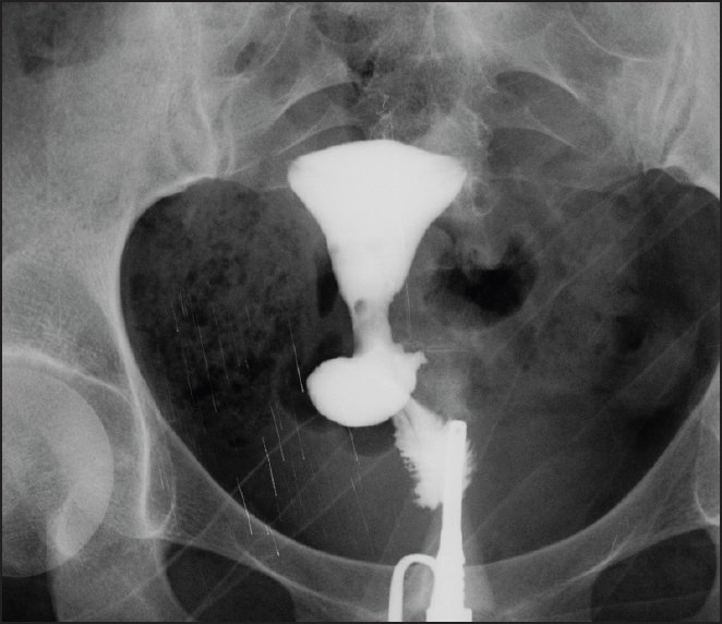
Hysterosalpingography (HSG) was performed during the proliferative phase and the shape of the endometrial cavity was normal. Bilateral tubal occlusion and saccular accumulation of contrast medium at 1/3 proximal level of cervix were also observed. A filling defect at the isthmus level measuring 3 × 4 mm was estimated to be an air bubble [Figure 1]