Surgeons have used robots operated by joysticks for more than a decade, and teams have shown that tiny robots can be steered through the body by external forces such as magnetism.
Now, bioengineers led by Harvard Medical School and Boston Children’s Hospital describe a robotic catheter that can navigate autonomously—the surgical equivalent of a self-driving car.
As reported April 24 in Science Robotics , the catheter found its way along the walls of a beating, blood-filled heart to a leaky valve in an animal model, without a surgeon’s involvement.
Senior investigator Pierre Dupont, professor of surgery at HMS and chief of pediatric cardiac bioengineering at Boston Children’s, believes this is the first instance of autonomous navigation inside an organism.
If the robotic catheter or future iterations prove safe and effective in humans, the device could eliminate the need for fluoroscopic imaging in these types of valve repairs, which exposes patients to ionizing radiation, Dupont said.
The autonomous technology also could provide guidance during particularly difficult or unusual cases, assist in operations in parts of the world that lack highly experienced surgeons, reduce learning curves for clinicians adopting new minimally invasive techniques, and offer benefits beyond navigation, such as conducting routine heart-mapping tasks.
Dupont hopes the self-driving robots will one day free surgeons to focus on the most difficult maneuvers, ultimately improving patient outcomes.
Consider the analogy of a fighter pilot and a fighter plane, he said. “The fighter plane takes on the routine tasks like flying the plane, so the pilot can focus on the higher-level tasks of the mission.”
Touch-guided vision, informed by AI
The catheter navigated using an optical touch sensor developed in Dupont’s lab, informed by a map of the cardiac anatomy and preoperative scans.
The sensor uses artificial intelligence and image processing algorithms to enable the catheter to figure out where it is in the heart and where it needs to go.
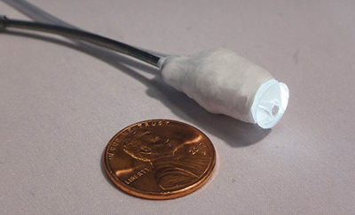
Catheter shown next to a penny for scale. Image: Margherita Mencattelli
For the demo, the team performed a technically demanding procedure known as paravalvular aortic leak closure, which repairs replacement heart valves that have begun leaking around the edges. (The team constructed its own valves for the experiments.) Once the robotic catheter reached the leak location, an experienced cardiac surgeon took control and inserted a plug to close the leak.
In repeated trials in swine, the robotic catheter successfully navigated to heart valve leaks in roughly the same amount of time as a surgeon using either a hand tool or a joystick-controlled robot.
Biologically inspired navigation
Through a navigational technique called wall following, the catheter’s sensor sampled its environment at regular intervals, in much the way insect antennae or rodent whiskers sample their surroundings to build mental maps of unfamiliar, dark environments.
The sensor told the catheter through images from a tip-mounted camera whether it was touching blood, the heart wall or a valve, as well as how hard it was pressing, to keep it from damaging the beating heart.