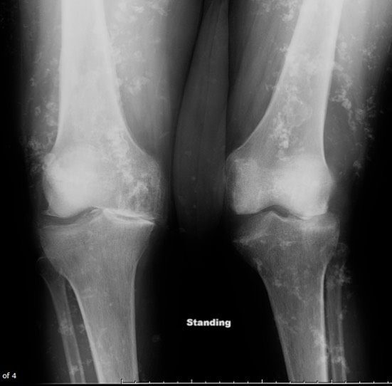
Soft Tissue Calcifications (STC)! STC are commonly encountered in all types of imaging studies and may often cause confusion. In general terms, STC are a consequence of either prior inflammation or trauma with tissue necrosis (dystrophic) or related to imbalances in calcium metabolism (metastatic). Thankfully, 95-98% of all the calcification we see on imaging are dystrophic and related to prior tissue necrosis including sequelae from prior trauma, systemic autoimmune inflammatory conditions, tumors, infections or ischemia. Not all dystrophic calcifications look the same, but we can differentiate some of them by location, distribution or clinical history. For example, diffuse soft tissue calcifications, like in this case, point to a systemic condition like dermatomyositis or generalized infections (parasites). Focal muscle calcification after trauma is likely sequelae from myonecrosis. Periarticular distribution of calcifications may suggest an inflammatory arthropathy. Metastatic calcifications are uncommon and related to disorders of calcium metabolism such as kidney disease, hyperparathyroidism or sarcoid, among others. So, if you see a soft tissue calcification and patient has normal calcium metabolism, try to direct your efforts in figuring out what caused soft tissue necrosis, which will point you to the most likely diagnosis. This is a case of Dermatomyositis