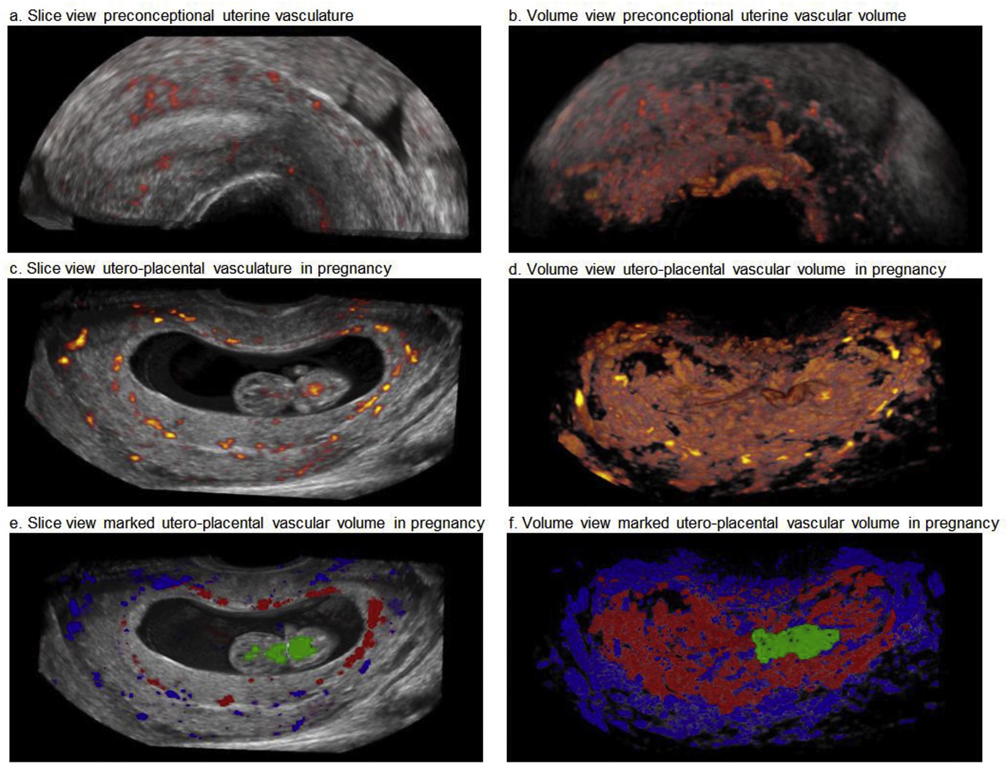
Fig. 1. Three-dimensional power Doppler ultrasound images of utero-placental vascular volumes preconceptional and in pregnancy at 9 weeks GA visualized by Virtual Reality (VR).
a: Slice view of a midsagittal uterine section showing the preconceptional uterine vascular volume (UVV, orange), with surrounding grey values representing the uterine tissue.
b: Volume view of figure 1a showing only the preconceptional uterine vascular volume (UVV, orange), after setting a threshold for grey values.
c: Slice view of a total obtained utero-placental vascular volume in pregnancy, with surrounding grey values representing the uterine tissue.
d: Volume view of figure 1c showing the total obtained utero-placental vascular volume in pregnancy, after setting a threshold for grey values.
e: Slice view of a total obtained utero-placental vascular volume in pregnancy, with surrounding grey values representing the uterine tissue and marked vessel subtypes (uterine vessels (blue); placental vessels (PVV, red); embryonic vessels (EVV, green).
f: Volume view of figure 1e, showing marked vessel subtypes (UVV (blue); placental vascular volume (PVV, red); embryonic vascular volume (EVV, green)).