Malakoplakia:
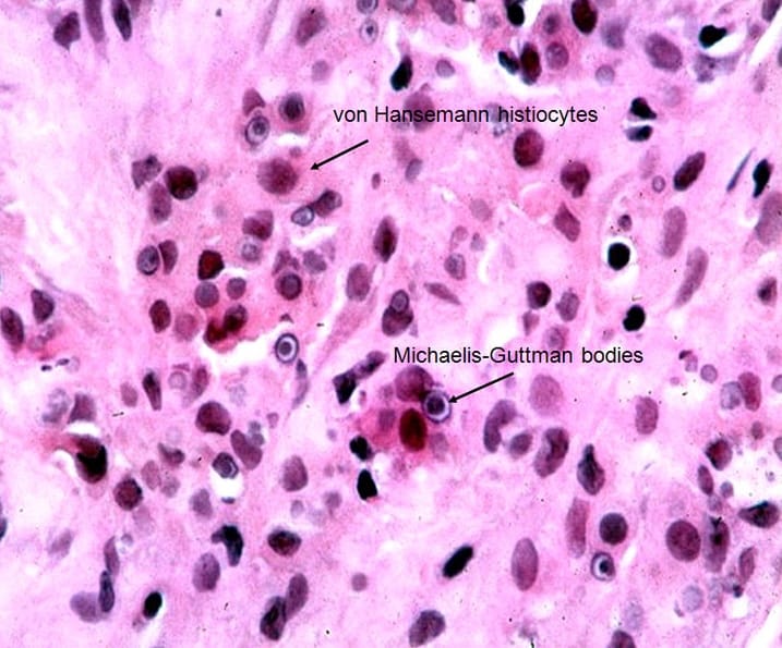
This stunning image of von Kossa stained section of bladder showing von Hansemann histiocytes and Michaelis- Guttman bodies!!! ![]()
![]()
![]()
![]()
![]()
![]()
![]()
Most often seen in women (75%) and peak at 50’s to 70’s.!Due to defective bactericidal capacity of histiocytes, as a result, ingested bacteria become calcified and laminated forming distinctive inclusions.
Usually caused by Escherichia coli and other gram-negative bacteria. Most common in bladder trigone, and may occur in other parts of GU tract. By cystoscopy, visible as single or multiple soft yellow or yellow-brown mucosal plaques (“malakos” = soft; “plakus” = plaques).
Histology:
Accumulation of histiocytes with pale pink foamy cytoplasm (von Hansemann histiocytes)
Diagnostic feature is the presence of intracytoplasmic round concentric basophilic inclusions (Michaelis-Guttman bodies).
Michaelis-Guttman bodies can be highlighted by PAS, von Kossa or iron stains.