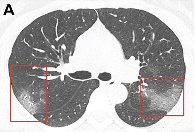
COVID 19 Pneumonia: Typical CT findings included bilateral pulmonary parenchymal ground-glass and consolidative pulmonary opacities, sometimes with a rounded morphology and a peripheral lung distribution. Notably, lung cavitation, discrete pulmonary nodules, pleural effusions, and lymphadenopathy were absent. Follow-up imaging in a subset of patients during the study time window often demonstrated mild or moderate progression of disease, as manifested by increasing extent and density of lung opacities.
CT is more sensitive than PCR