Introduction
Multiple self-healing palmoplantar carcinoma (MSPC) is a rare, autosomal dominant genetic condition caused by a gain-of-function mutation in the NLRP1 gene, which primarily affects epithelium lacking hair follicles, such as the palms and soles, leading to multiple keratoacanthomas (KAs). MSPC belongs to a family of syndromes that manifest with multiple KAs, including Muir-Torre syndrome, Witten-Zak syndrome, Grzybowski syndrome, and multiple self-healing squamous epithelioma.[1](javascript:void(0)![]() Here we report 2 cases of MSPC involving a mother and daughter with concomitant hidradenitis suppurativa (HS).
Here we report 2 cases of MSPC involving a mother and daughter with concomitant hidradenitis suppurativa (HS).
Case report
A mother and daughter presented to the dermatology clinic with similar hyperkeratotic papules on the palms, soles, and extremities along with cystic nodules in the groin. The daughter was a 13-year-old girl with a history of asthma and multiple laryngeal papillomas with multiple skin lesions on her upper and lower extremities that were treated as common warts with cryotherapy for several years. The mother of the patient noted that papules initially appeared on the patient’s palms and soles around 2 years of age and underwent periods of growth and regression. Additionally notable was a history of odontohypophosphatasia with early tooth loss beginning at age 6 with complete loss of deciduous teeth by age 10 along with current loosening of permanent teeth. To date, she has not had conjunctival lesions but does have a history of myopia.
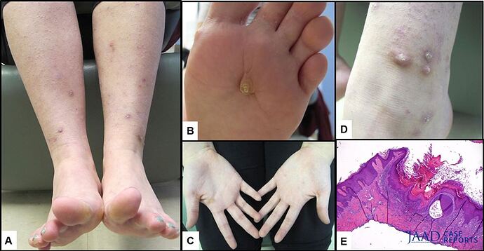
Physical examination found hyperkeratotic crusted papules and plaques on the palms, soles, and legs along with scattered hyperpigmented macules and patches in areas of previous lesions (Fig 1, A , B , and D ). Additionally, involving the bilateral upper thighs and inguinal creases were pink-to-brown cystic nodules consistent with HS (Hurley stage 1). Keratoderma was present on her palms and soles bilaterally (Fig 1, C ). Shave biopsy of a lesion on her left ankle found histology consistent with a KA (Fig 1, E ), and the lesion was negative for high-risk human papillomavirus infection. She underwent whole genome sequencing which found a significant pathologic mutation in the NLRP1 gene on chromosome 17p13 with a c.197 C>T (p.A66V) variant. Of note, this genetic change was also identified in the patient’s mother, maternal aunt, and maternal grandmother. Further workup included an interleukin (IL)-1 level, which was within normal limits.
To address the KA lesions, she was started on tretinoin 0.05% cream and oral niacinamide (500 mg twice a day) supplementation. Furthermore, to treat the HS, she was given clindamycin 1% topical solution and Hibiclens 4% topical solution. At 6-month follow-up, she had minimal improvement with either condition so her topical retinoid was switched to tazarotene and her HS was more aggressively managed with intralesional triamcinolone injections and oral doxycycline with the consideration to start isotretinoin in the near future. To date, squamous cell carcinoma has not been diagnosed.
The mother was a 45-year-old woman who presented with multiple self-healing lesions on her lower legs and feet that started during childhood. She also had history of odontohypophosphatasia with early loss of her deciduous teeth along with lack of classic ocular findings. Physical examination found multiple hyperkeratotic firm papules on the legs (Fig 2, A ). Involving the groin were small cystic red-to-brown papules consistent with HS (Hurley stage 1). Yellow keratoderma was present on the pressure points of the palms and soles bilaterally (Fig 2, B ). Biopsy of 2 characteristic lesions on the left and right legs were consistent with invasive squamous cell carcinoma. After both lesions were excised, the patient was started on acitretin (10 mg/d) along with oral niacinamide (500 mg twice a day) supplementation. At 4-month follow-up, the KA and HS lesions completely resolved. The treatment regimen was well tolerated by the patient and subsequently continued. Unfortunately, breast cancer was recently diagnosed in this patient.
Discussion
The ability of the integumentary system to respond appropriately and judiciously is crucial for the health and survival of the individual. One major pathway for inflammation and host defense involves the previously described inflammasome complexes. These complexes are assembled in response to extracellular danger-associated molecular patterns (DAMPs). DAMPs consist of a wide variety of molecular patterns including bacterial toxins, free adenosine triphosphate, and electrolyte imbalances. The DAMPs interact with Toll-like receptors and C-type lectin receptors in the cell membrane. These receptors in turn activate different pattern recognition receptors (PRRs) in the cytoplasm. The human PRRs include NLRP1, NLRP3, NLRC4, AIM1, and pyrin. Once mobilized, the PRRs assemble the macromolecular inflammasome complexes and initiate cleavage of pro–caspase-1 into caspase-1, promoting apoptosis and the release of inflammatory extracellular cytokines such as IL-18 and IL-1 β . Assembly of the inflammasome complexes can be initiated by any of the PRRs.[2](javascript:void(0);), [3](javascript:void(0)![]() Therefore, mutations in genes that encode any of these PRRs can lead to aberrant inflammasome assembly and function. In fact, Zhong et al[1](javascript:void(0)
Therefore, mutations in genes that encode any of these PRRs can lead to aberrant inflammasome assembly and function. In fact, Zhong et al[1](javascript:void(0)![]() described increased inflammasome activity in mutant human cells that had a gain-of-function NLRP1 mutation compared with wild-type cells in vitro.
described increased inflammasome activity in mutant human cells that had a gain-of-function NLRP1 mutation compared with wild-type cells in vitro.
Changes in the NLRP1 gene can result in 2 distinct clinical conditions: multiple self-healing palmoplantar carcinoma and familial keratosis lichenoides chronica. MSPC is associated with recurrent KAs on the palms and soles along with corneal intraepithelial dyskeratosis, corneal opacification with dyskeratosis, palmoplantar hyperkeratosis, dyshidrosis, maxillary decalcification with tooth loss, and hyperkeratosis pilaris. Mamaï et al[4](javascript:void(0)![]() initially described the autosomal dominant transmission of MSPC through 27 individuals of a Tunisian family. They found the average age of onset to be 8.8 years in those studied who were younger than 25 years with the life cycle of the KAs lasting 3 months on average. By comparing the genotypes of the affected and unaffected family members, they were able to localize the aberrant gene to chromosome 17.p13.3-12. Clinically, the lesions appear similar to KAs, beginning as a papule with gradual growth that eventually ulcerates and spontaneously regresses. However, from a histologic standpoint these lesions more closely resemble squamous cell carcinomas.[4](javascript:void(0);), [5](javascript:void(0)
initially described the autosomal dominant transmission of MSPC through 27 individuals of a Tunisian family. They found the average age of onset to be 8.8 years in those studied who were younger than 25 years with the life cycle of the KAs lasting 3 months on average. By comparing the genotypes of the affected and unaffected family members, they were able to localize the aberrant gene to chromosome 17.p13.3-12. Clinically, the lesions appear similar to KAs, beginning as a papule with gradual growth that eventually ulcerates and spontaneously regresses. However, from a histologic standpoint these lesions more closely resemble squamous cell carcinomas.[4](javascript:void(0);), [5](javascript:void(0)![]()
The simultaneous manifestation of MSPC and HS in a patient appears to follow a genetic pattern and has yet to be described. With the confirmed NLRP1 gain-of-function mutation in the patient, inflammasome hyperactivity led to the development of multiple KAs. Could this inappropriate inflammasome activation contribute to the development of her inflamed HS lesions? Although follicular plugging and mechanical friction are suggested to contribute to the formation of the furuncles, the inflammatory response from the host propagates it. It is likely that the inappropriate elevation of cytokines, specifically IL-1 β , from the NLRP1 mutation contribute significantly to the propagation of her HS. Previous studies found elevated levels of IL-1 β in patients with HS that correlate with disease severity, suggesting a strong relationship between the disease and a systemic pro-inflammatory state.[6](javascript:void(0);), [7](javascript:void(0)![]() However, more extensive investigation will have to be done to evaluate the relationship between these 2 complex inflammatory diseases.
However, more extensive investigation will have to be done to evaluate the relationship between these 2 complex inflammatory diseases.
