On this page:
Superficial MusclesDeep MusclesMuscles of the shoulder & armShoulder Movements
Shoulder Muscles: The shoulder muscles are associated with movements of the upper limb. The shoulder muscles produce the characteristic shape of the shoulder and can be classified into two groups:
Extrinsic shoulder muscles – arise from the torso, and inserts to the clavicle, scapula or humerus).
Intrinsic shoulder muscles – arise from the scapula and/or the clavicle, and inserts to the humerus.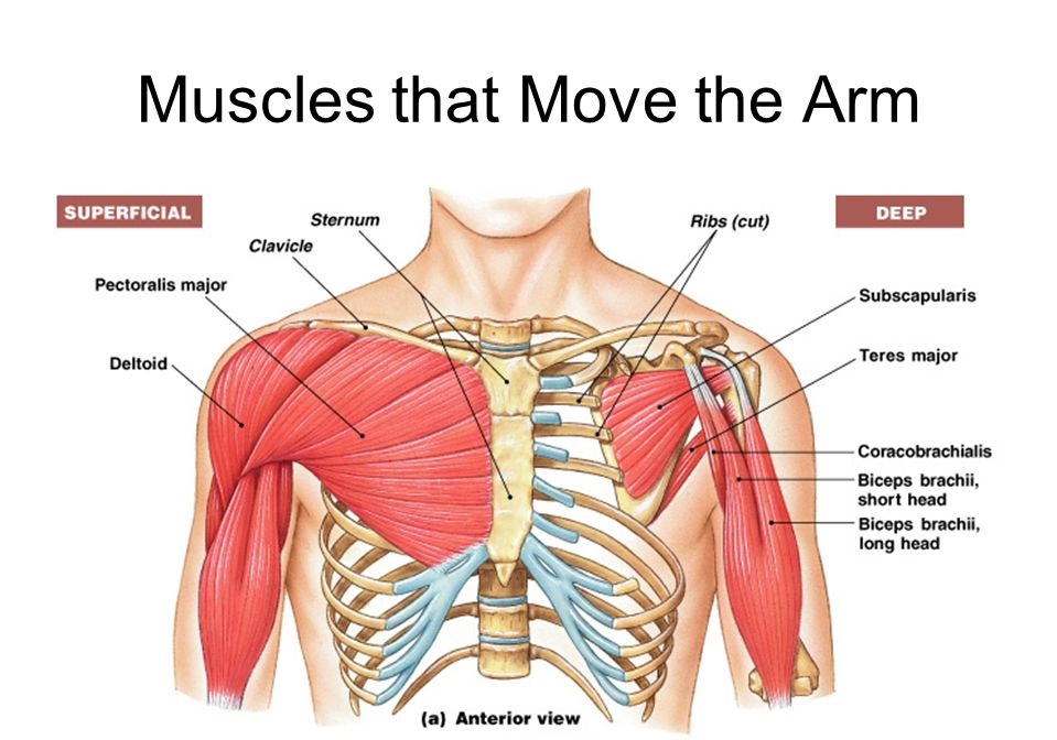 The shoulder muscles can be separated into three important groups:
- Superficial muscles (Extrinsic)
- Deep muscles (Intrinsic)
- Muscles of the shoulder & arm
Superficial Muscles
Pectoralis Major
Trapezius
Latissimus dorsi
Deltoid
Pectoralis Major Muscle: The pectoralis major is a thick, fan-shaped muscle, located at the chest. It makes up the bulk of the chest muscles and rests under the breast.
Origin: It has two head.
Clavicular head:
Anterior surface of medial half of clavicle
Sternal head:
Anterior surface of manubrium and sternum up to sixth costal cartilages
2nd to 6th costal cartilages
Aponeurosis of the external oblique muscle of the abdomen
Insertion: Lateral lip of bicipital groove of humerus
Nerve supply: Medial and lateral pectoral nerves
Action:
Adduction and medial rotation of the shoulder
Clavicular part produces flexion of the arm
Sternal part is used in
Extension of the flexed arm against resistance
Climbing
Trapezius: The trapezius is a large, flat, superficial muscle lengthening from the cervical to thoracic area on the posterior aspect of the neck and trunk. The muscle is split into three parts: superior, inferior, and middle part.
Origin:
Occipital protuberance
Ligamentum nuchae
Spine of the 7th cervical vertebra
Spine of all thoracic vertebra
Insertion:
Upper fibers of trapezius into posterior border of the lateral third of the clavicle
Middle fibers into the medial margin of the acromion and upper lip of the crest of the spine of the scapula
Lower fibers on the deltoid tubercle
Nerve supply: Spinal part of the accessory nerve is the motor, branches from C3, C4 are proprioceptive
Action:
Upper fibers elevate the scapula
Middle fibers retract the scapula
Upper and lower fibers rotate the scapula forwards
Latissimus Dorsi: The latissimus dorsi a large, flat muscle on the back and, behind the arm, and is notably covered by the trapezius on the back near the midline. The latissimus dorsi is the longest muscle in the upper body.
Origin:
Posterior one-third of the outer lip of iliac crest
Posterior layer of lumbar fascia
Spines of the T7-T12 vertebra
Lower four ribs
Inferior angle of the scapula
Insertion: Bicipital groove of the humerus
Nerve supply: Thoracodorsal nerve ( nerve to latissimus dorsi)
Action:
Adduction, extension and medial rotation of the shoulder
Deltoid: The Deltoid is a large triangular shaped muscle which extends over the glenohumeral joint and which provides the shoulder its rounded contour. The Deltoid comprises 3 distinct pieces each of which offers a different movement of the glenohumeral joint, usually identified the anterior, mid and posterior heads.
Origin: Lateral third of clavicle, acromion, and spine of scapula
Insertion: Deltoid tuberosity of the humerus
Function: Anterior part of the deltoid: flexes and medially rotates arm; Middle partof the deltoid: abducts arm; Posterior part of the deltoid: extends and laterally rotates the arm
Nerve Supply: Axillary nerve (C5 and C6)
Deep Muscles
Pectoralis minor
Subclavius
Levator Scapulae
Rhomboid major and minor
Teres major
Serratus anterior
The rotator cuff muscles
Supraspinatus
Infraspinatus
Teres minor
Subscapularis
Pectoralis Minor Muscle: The pectoralis minor is a thin, triangular muscle, located at the upper part of the chest, under the pectoralis major.
Origin:
3,4,5 ribs, near the costochondral junction
Intervening fascia covering external intercostal muscles
Insertion: Medial border and the upper surface of the coracoid process of scapula
Nerve supply: Medial and lateral pectoral nerves
Action:
Drows the scapula forward ( with serratus anterior)
Depresses the point of the shoulder
Helps in forced inspiration
Subclavius: The subclavius muscle is small triangular, located within the clavicle and the first rib. Along with the pectoralis minor and pectoralis major muscles, the muscle of the subclavius makes up the anterior wall of the axilla.
Origin: Subclavius originated from 1st rib at the costochondral junction
Insertion: It inserts to the subclavian groove in the middle one-third of clavicle
Nerve supply: Nerve to subclavius from upper trunk of brachial plexus
Action: Steadies the clavicle during movements of the shoulder
Serratus Anterior: The serratus anterior originates on the surface of the 1st to 8th ribs at the side of the chest and attaches with the entire anterior length of the medial border of the scapula.
Origin: Upper eight ribs
Insertion:Costal surface of the scapula along its medial border
Nerve supply: Long thoracic nerve
Action:
Abductor of the shoulder
Protractor of the scapula
Levator Scapulae: The levator scapulae is located at the back and side of the neck. Its main function is to lift the scapula.
Origin: Transverse process of first four cervical vertebra
Insertion: Superior angle and upper part of medial border of scapula
Nerve supply: Dorsal scapular nerve
Action: Elevation of the scapula
Rhomboideus Minor: The rhomboid minor is located on the back that attaches the scapula to the vertebrae of the spinal column.
Origin:
Lower part of ligamentum nuchae
Spine of C7 and T1 vertebra
Insertion: Upper 1/3rd of medial border of scapula
Nerve supply: Dorsal scapular nerve
Action: Retraction of the scapula
Rhomboideus Major: The rhomboid major is located on the back that connects the scapula among the vertebrae of the spinal column.
Origin: Spine of the T2-T5 vertebra
Insertion: Medial border of scapula below the root of the spine
Nerve supply: Dorsal scapular nerve
Action: Retraction of the scapula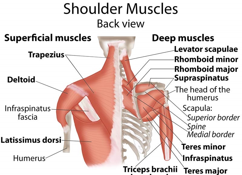
Supraspinatus: The supraspinatus is located upper back that runs from the supraspinatus fossa superior portion of the scapula to the greater tubercle of the humerus. The supraspinatus is one of the four rotator cuff muscles.
Origin: Supraspinatus originated from Supraspinous fossa of the scapula
Insertion: It inserts to the Superior facet on greater tuberosity of humerus
Function: Supraspinatus initiates and helps deltoid in abduction of the arm and acts with other rotator cuff muscles
Nerve Supply: Suprascapular nerve (C4, C5, and C6)
Infraspinatus: The infraspinatus is a triangular muscle, which holds the chief part of the infraspinatous fossa. The infraspinatus is one of the four rotator cuff muscles.
Origin: Infraspinatus originated from Infraspinous fossa of the scapula
Insertion: It inserts to the Middle facet on greater tuberosity of humerus
Function: Laterally rotate arm; helps to hold humeral head in the glenoid cavity of the scapula
Nerve Supply: Suprascapular nerve (C5 and C6)
Teres Minor: The teres minor is a narrow, elongated muscle of the rotator cuff. The teres minor muscle originates from the lateral border of the scapula and inserts to the greater tubercle of the humerus including the posterior surface of the joint capsule.
Origin: Teres Minor originated from Superior part of the lateral border of scapula
Insertion: It inserts to the Inferior facet on greater tuberosity of humerus
Function: Laterally rotate arm; helps to hold humeral head in the glenoid cavity of the scapula
Nerve Supply: Axillary nerve (C5 and C6)
Subscapularis: The subscapularis which fills the subscapular fossa and inserts into the lesser tubercle of the humerus and the front part of the capsule of the shoulder joint. The Subscapularis is one of the four rotator cuff muscles.
Origin: Subscapularis originated from Subscapular fossa of the scapula
Insertion: It inserts to the Lesser tuberosity of humerus
Function: Medially rotates the arm and adducts it; helps to hold humeral head in the glenoid cavity of the scapula
Nerve SUPPLY: Upper and lower subscapular nerves (C5, C6, and C7)
Muscles of the shoulder & arm
Biceps brachii
Coracobrachialis
Triceps brachii
Biceps Brachii: The biceps brachii is a two-headed muscle that lies on the upper arm connecting the shoulder and the elbow.
Origin: Short head of the Biceps Brachii: the tip of coracoid process of scapula; Long head of the Biceps Brachii: supraglenoid tubercle of scapula
Insertion: It inserts to the Tuberosity of radius and fascia of forearm via the bicipital aponeurosis
Function: Supinates forearm and, when it is supine, flexes the forearm
Nerve Supply: Musculocutaneous nerve (C5 and C6 )
Coracobrachialis: The coracobrachialis is situated at the upper and medial part of the arm.
Origin: Coracobrachialis originates from Tip of coracoid process of scapula
Insertion: It inserts to the Middle third of the medial surface of the humerus
Function: Helps to flex and adduct the arm
Nerve Supply: Musculocutaneous nerve (C5, C6, and C7)
Triceps Brachii: The triceps brachii is a large muscle on the back of the upper limb. The triceps brachii is principally responsible for extension of the elbow joint.
Origin: Long head of the Triceps Brachii: infraglenoid tubercle of scapula; Lateral head of the Triceps Brachii: posterior surface of the humerus, superior to radial groove; Medial head of the Triceps Brachii: posterior surface of the humerus, inferior to radial groove
Insertion: It inserts to the Proximal end of olecranon process of ulna and fascia of forearm
Function: Triceps Brachii main function is extensor of the forearm; long head steadies head of abducted of the humerus
Nerve Supply: Radial nerve (C6, C7, and C8)
Shoulder Movements
Shoulder Flexion: The straight arm is raised in front of the body, with the palm down, as high as possible.
Shoulder Flexion Muscles: Pectoralis major, anterior deltoid, and coracobrachialis. Biceps brachii weakly assists in forwarding flexion.
Shoulder Extension: Shoulder extension is when you lift your arms behind your back in the sagittal plane.
Shoulder Extension Muscles: Posterior fibers of the deltoid, latissimus dorsi, and teres major.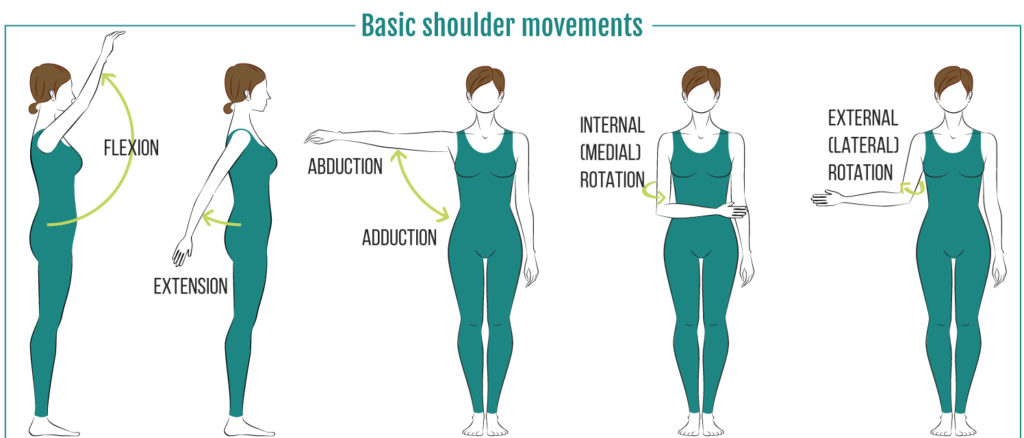
Shoulder Abduction: The straight arm is raised at the side, with the palm down, as high as possible.
Shoulder Abduction Muscles: The first 0-15 degrees of the shoulder abduction is produced by the supraspinatus. The middle fibers of the deltoid are engaged for the next 15-90 degrees. Past 90 degrees– that is carried out by the trapezius and serratus anterior.
Shoulder Adduction: Shoulder adduction is a medial movement at the shoulder (glenohumeral) joint – moving the upper arm down to the side towards the body.
Shoulder Adduction Muscles: Pectoralis major, latissimus dorsi, and teres major.
Shoulder Internal Rotation: The arm is put behind the back with the elbow bent. The person reaches as far up the back as possible. This distance is measured from a specific point on the spine.
Shoulder Internal Rotation Muscles: Subscapularis, pectoralis major, latissimus dorsi, teres major and anterior deltoid.
Shoulder External Rotation: The elbows are held by the sides of the body, bent at 90o with palms facing each other. Then, keeping the elbows in contact with the body, the hands are spread outwards as far as possible.
Shoulder External Rotation Muscles: Infraspinatus and teres minor.
You Might Also Like:
Frozen Shoulder (Adhesive Capsulitis): Symptoms, Causes, Treatment & Exercises
Shoulder Impingement Syndrome: Symptoms, Causes, Diagnosis & Treatment
Shoulder Dislocation : Causes,Types,Symptoms & Treatment
Rotator Cuff vs Frozen Shoulder
Glycemic Index: High & Low Glycemic Foods
Tags :